From Modic changes and clinical phenotype to targeted basivertebral nerve treatment
Chronic low back pain is not a single clinical entity. Different anatomical structures may act as pain generators, and identifying the predominant source of pain is essential for selecting an appropriate treatment strategy.
One distinct phenotype is vertebrogenic low back pain, which originates from pathology of the vertebral endplates and adjacent vertebral bone marrow. On MRI, this phenotype is most commonly associated with Modic type 1 or type 2 changes. The basivertebral nerve (BVN), which innervates the vertebral body and endplate region, provides a potential therapeutic target for appropriately selected patients. [1]
The Healthy Vertebra: Understanding the Normal Anatomy
The vertebral body provides structural support and distributes mechanical loads transmitted through the intervertebral disc. At its superior and inferior aspects, the vertebral endplates form the interface between the disc and vertebral body and play an important role in load transmission and disc–vertebral body homeostasis. [1]
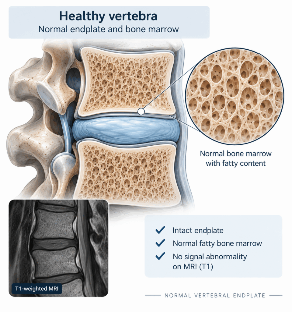
The vertebral body also contains an intraosseous sensory nerve network. The basivertebral nerve enters the vertebral body through the basivertebral foramen and branches toward the superior and inferior endplate regions. These neural structures are clinically relevant because damaged vertebral endplates can become a source of nociceptive input. [1]
In a healthy vertebral segment, the adjacent bone marrow and endplates do not demonstrate the characteristic MRI signal abnormalities associated with Modic changes.
Understanding this normal anatomy provides the basis for understanding what changes when vertebral endplate pathology develops.
Modic changes – an important MRI phenotype
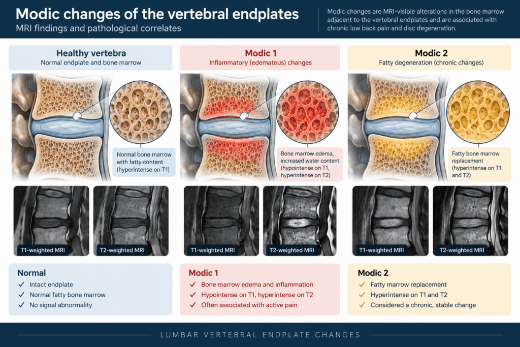
Modic changes are MRI-detectable signal alterations involving the vertebral bone marrow adjacent to the endplates. They were originally classified into three types based on their characteristic MRI appearance and underlying tissue changes. [2,3]
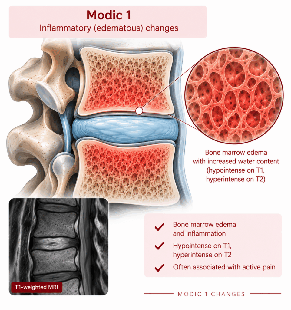
Modic type 1
Modic type 1 changes are generally considered the more biologically active phenotype and are associated with bone marrow edema, fibrovascular tissue and inflammatory changes.
Typical MRI appearance:
- T1-weighted: hypointense
- T2-weighted: hyperintense
Histologically, these changes correspond to edema and increased vascularity within the vertebral marrow. [2,3]
Modic type 2
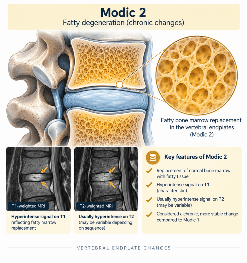
Modic type 2 changes represent a different pattern of vertebral marrow remodeling, characterized predominantly by fatty replacement of normal bone marrow.
Typical MRI appearance:
- T1-weighted: hyperintense
- T2-weighted: iso- or hyperintense depending on the imaging sequence
On fat-suppressed sequences, the fatty signal is typically suppressed. Mixed Modic type 1/2 patterns are also frequently encountered. [3]
Modic type 3
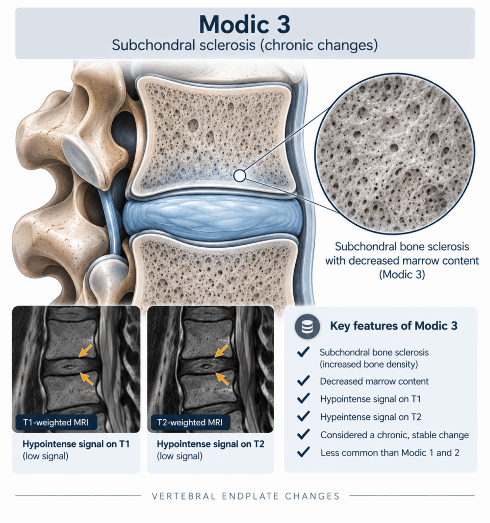
Modic type 3 changes are much less Modic type 3 changes are less common and are associated with subchondral sclerosis.
Typical MRI appearance:
- T1-weighted: hypointense
- T2-weighted: hypointense
Although Modic changes may be associated with low back pain, they are imaging findings rather than a diagnosis by themselves. Their clinical significance must be interpreted together with the patient’s symptoms, examination and differential diagnosis. [3,4]
Recognising the Vertebrogenic Pain Phenotype
Vertebrogenic pain typically presents as chronic, predominantly axial low back pain. Patients may describe deep, central or paracentral lumbar pain that can be aggravated by sitting, bending forward, loading or repetitive spinal activities. [1]
The clinical history should address:
- duration and progression of symptoms
- location and character of pain
- aggravating and relieving factors
- functional limitations
- previous conservative treatment
- neurological symptoms
- previous spinal interventions
- possible alternative pain generators
Physical examination may reproduce the patient’s familiar pain with flexion-based loading or stress on the anterior spinal column. However, no single symptom or examination finding is sufficient to establish a vertebrogenic diagnosis. [1]
Radicular pain, neurogenic claudication, facet-mediated pain, sacroiliac joint pain, hip pathology and other spinal or non-spinal conditions should therefore be considered as part of the differential diagnosis.
The diagnosis ultimately depends on concordance between the clinical phenotype and appropriate imaging findings, together with exclusion of other dominant pain generators. [1,5]
Who Is a Candidate for BVNA?
A typical candidate for basivertebral nerve ablation has:
- chronic predominantly axial low back pain, generally lasting more than six months
- persistent symptoms despite appropriate conservative treatment
- MRI evidence of Modic type 1 and/or type 2 changes at a clinically concordant level
- significant pain-related functional limitation
- no dominant alternative pain generator explaining the clinical presentation
- no relevant contraindication to the procedure [1,5]
Modic changes alone are not an indication for BVNA. The indication is based on the combination of a compatible clinical phenotype, concordant imaging and appropriate exclusion of competing pain generators.
Appropriate patient selection is therefore at least as important as the technical execution of the procedure.
Why Target the Basivertebral Nerve?
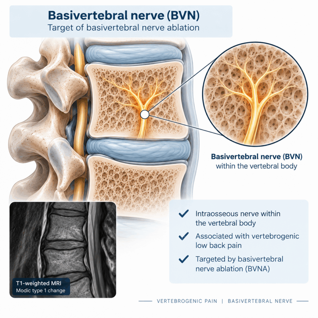
The vertebral endplates are richly innervated, and the basivertebral nerve provides an important sensory pathway from the vertebral body and endplate region. [1]
Endplate damage may be accompanied by inflammatory and neurogenic changes, including increased nociceptive signalling within the vertebral body. This provides the biological rationale for targeting the BVN in patients with a vertebrogenic pain phenotype. [1,6]
Basivertebral nerve ablation (BVNA) aims to interrupt this intraosseous pain pathway by selectively treating the basivertebral nerve within the vertebral body.
Unlike procedures directed primarily at the intervertebral disc, facet joints or peripheral nerves, BVNA targets a specific intraosseous neural structure associated with vertebral endplate pain.
Straight-Probe BVNA: Key Procedural Steps
BVNA is performed under image guidance using an intraosseous approach to reach the basivertebral nerve within the vertebral body. The essential procedural sequence can be summarised as follows:
1. Pre-procedural assessment
Confirm the clinical phenotype, review the MRI and establish the vertebral level or levels that are clinically and radiographically concordant.
2. Patient positioning
The patient is positioned prone, with appropriate support and attention to spinal alignment and pressure points.
3. Fluoroscopic planning
AP and lateral fluoroscopic views are used to identify the target vertebral body, pedicle and planned access trajectory.
4. Transpedicular access
A controlled transpedicular pathway is established into the vertebral body.
5. Intraosseous targeting
The working pathway is advanced toward the region of the basivertebral nerve.
6. Straight-probe positioning and ablation
The straight probe is positioned at the intended treatment site and radiofrequency energy is delivered according to the specific device and institutional protocol.
7. Completion and post-procedural care
The access is withdrawn and the patient is monitored according to the clinical protocol, including assessment of neurological status and post-procedural symptoms.
The exact instruments, access trajectory, probe configuration and RF parameters may vary according to the equipment and procedural protocol used. Detailed technical training under appropriate supervision is therefore essential.
What Does the Evidence Show?
The clinical evidence supporting BVNA has expanded substantially over the past decade. Randomized trials and prospective studies have demonstrated clinically meaningful improvements in pain and disability in appropriately selected patients with vertebrogenic low back pain. The ASPN best-practice guidelines assign BVNA a Grade A recommendation with high certainty of substantial net benefit in appropriately selected patients. [1]
Long-term follow-up has also demonstrated durability of treatment effect. Five-year follow-up from a prospective randomized study showed sustained improvements in pain and function, supporting BVNA as a treatment option for a specific subgroup of patients with chronic vertebrogenic low back pain. [7]
A subsequent pooled five-year analysis of three prospective clinical trials similarly demonstrated sustained improvements in pain and disability, with a substantial proportion of patients reporting clinically meaningful improvement. [8]
These findings reinforce an important principle: BVNA should not be regarded as a treatment for MRI abnormalities alone. The clinical diagnosis and selection of the appropriate patient remain fundamental.
From Clinical Diagnosis to Hands-on Training
The transition from understanding vertebrogenic pain to performing BVNA safely requires more than theoretical knowledge. Precise understanding of vertebral anatomy, fluoroscopic landmarks, access trajectories and procedural technique is essential.
These principles form part of the educational programme of the Interventional Pain Academy by EuroPainClinics.
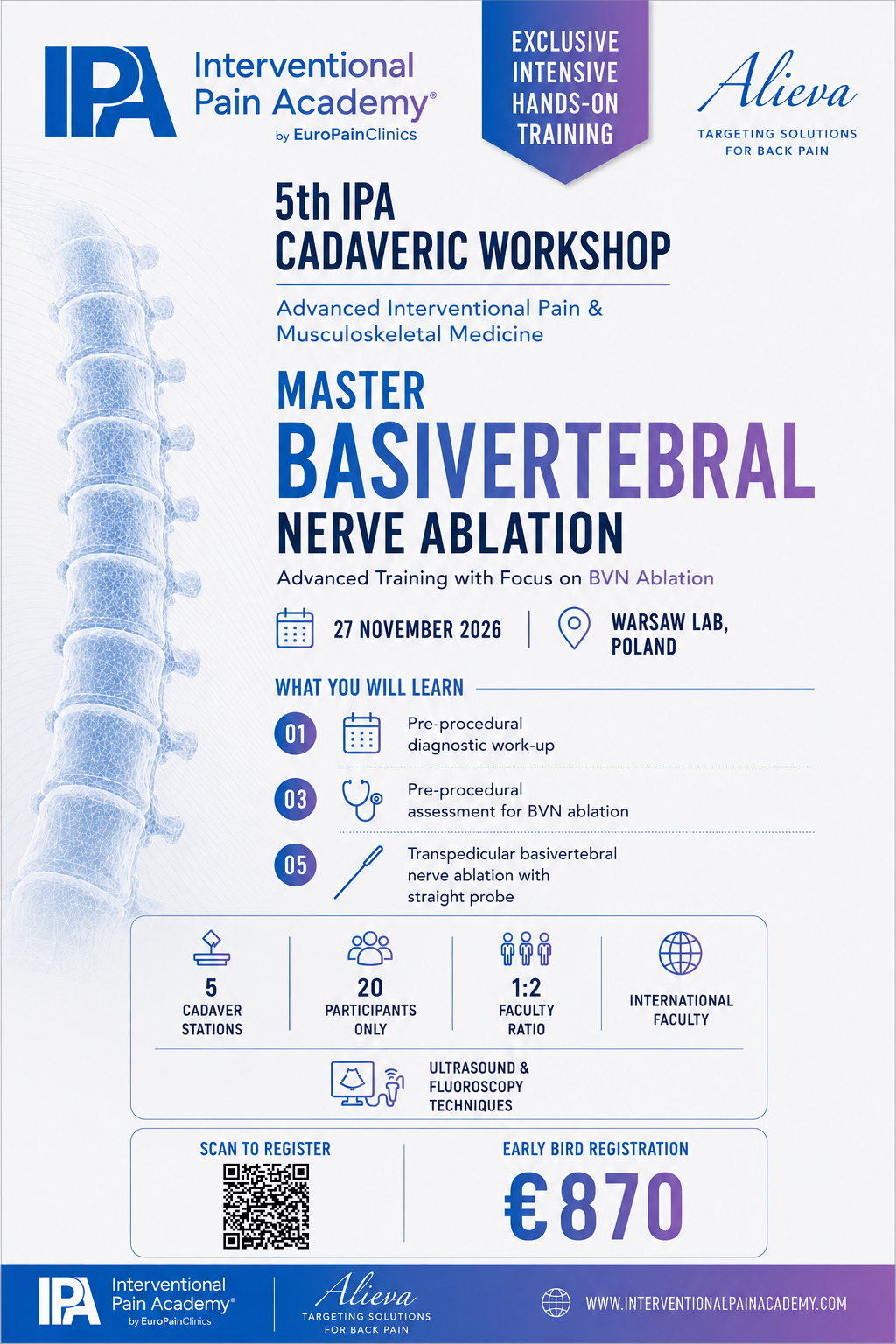
The 5th IPA Cadaveric Workshop – Warsaw 2026, taking place on 27 November 2026, includes dedicated training in pre-procedural assessment for basivertebral nerve ablation and transpedicular BVNA. The workshop combines lectures, interactive discussion and intensive cadaver-based hands-on training with an international faculty. [9]
A dedicated BVNA session will also be presented by Richard Harker, FRCS (Tr & Orth), UK, including his clinical experience and own clinical data.
For physicians interested in understanding the anatomy, patient selection and practical principles of BVNA, cadaveric training provides an opportunity to connect the clinical concept with the technical realities of the procedure.
Conclusion
Vertebrogenic low back pain represents a distinct clinical phenotype in which the vertebral endplate and adjacent bone marrow play a central role.
Recognising the difference between a healthy vertebral segment and Modic type 1 or type 2 changes provides an important foundation for understanding this condition. However, MRI findings must always be interpreted in the context of the patient’s history, clinical examination and differential diagnosis.
For appropriately selected patients, basivertebral nerve ablation provides a targeted approach to treating vertebrogenic pain by interrupting the intraosseous sensory pathway associated with the affected vertebral endplates.
Ultimately, successful BVNA begins well before the procedure itself:
The right patient.
The right diagnosis.
The right target.
The right technique.
The right patient. The right diagnosis. The right target. The right technique.
That combination forms the foundation of contemporary vertebrogenic pain management and represents the focus of the dedicated BVNA teaching and hands-on training at the 5th IPA Cadaveric Workshop – Warsaw 2026.
References:
- Sayed D, Naidu RK, Patel KV, et al. Best Practice Guidelines on the Diagnosis and Treatment of Vertebrogenic Pain with Basivertebral Nerve Ablation from the American Society of Pain and Neuroscience. J Pain Res.2022;15:2801-2819. doi:10.2147/JPR.S378544.
- Modic MT, Steinberg PM, Ross JS, Masaryk TJ, Carter JR. Degenerative disk disease: assessment of changes in vertebral body marrow with MR imaging. Radiology. 1988;166(1 Pt 1):193-199.
- Zhang YH, Zhao CQ, Jiang LS, Chen XD, Dai LY. Modic changes: a systematic review of the literature. Eur Spine J. 2008;17:1289-1299.
- Herlin C, Chica-Rios A, Kjaer P, et al. Modic changes—their associations with low back pain and activity limitation: a systematic literature review and meta-analysis. PLoS One. 2018;13(8):e0200677.
- Deer TR, Sayed D, Michels J, et al. The American Society of Pain and Neuroscience (ASPN) Evidence-Based Clinical Guideline of Interventional Treatments for Low Back Pain. J Pain Res. 2022;15:3729-3758.
- Khan M, et al. Basivertebral Nerve Ablation: Part 1—Anatomy, Pathophysiology, and Clinical Rationale. AJNR Am J Neuroradiol. 2026;47(6):1750.
- Fischgrund JS, Rhyne A, Macadaeg K, et al. Long-term outcomes following intraosseous basivertebral nerve ablation for the treatment of chronic low back pain: 5-year treatment arm results from a prospective randomized double-blind sham-controlled multicenter study. Eur Spine J. 2020;29(8):1925-1934.
- Khalil JG, et al. Intraosseous basivertebral nerve ablation: a 5-year pooled analysis from three prospective clinical trials. Spine J. 2025.
- Interventional Pain Academy. 5th IPA Cadaveric Workshop – Warsaw 2026. November 27, 2026. Interventional Pain Academy.
[1] ASPN Best Practice Guidelines — Full-text article – PMC
[3] Modic changes – systematic review — Full-text article – PMC
[4] Modic changes – systematic review and meta-analysis — Full-text article – PMC
[7] Five-year BVNA outcomes — PubMed
[8] Five-year pooled BVNA analysis — PubMed
[9] IPA Courses / Workshop — Interventional Pain Academy – Courses